
Back Pain: The Truth about MRI Scans
- In India, every 3 out of 10 individuals suffer from chronic back pain. And even that number seems insignificant, if you consider the fact that most of us personally know at least one person suffering from this condition.
Read on to find out.
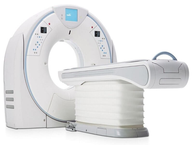
But first, when do you need an MRI, as opposed to a CT scan? An MRI focuses on soft tissues and muscles and helps diagnose related problems. A CT scan, on the other hand, is recommended if you have a bone fracture, or need to examine problems with the bone. This is the simplest and most important difference to remember.
When should you consider getting that MRI scan?
Of course, it goes without saying that you should only get an MRI scan after your doctor/ orthopaedic recommends it. But for your own knowledge, here’s when an MRI scan may be essential:
A CT scan, on the other hand, is recommended if you have a bone fracture, or need to examine problems with the bone.
1. If you have severe neck or back pain that does not go away
2. If you’re experiencing significant weakness, numbness, or changes in reflex that have developed rapidly (which could be due to nerve damage)
3. If you’re suffering from a sudden loss of muscle control or bodily functions, including loss of bowel/bladder control
4. If you’re not experiencing any of these symptoms, we recommend you adopt the wait and watch approach, while continuing your
non-invasive line of treatment (including exercises and over-the-counter pills) – given time, this should help your back pain get better or become more manageable.
Read this before getting an MRI scan
An MRI scan is crucial in many cases and can change your life for the better. However, at the same time, drawing a diagnosis from an MRI report is tricky. For example, your scan may show a lumbar disc herniation/disc degeneration, which sounds like a serious problem. However, studies show that approximately 30% of individuals in their 30s and 40s suffer from this condition, without even realizing it, because it doesn’t affect their daily life. Which means that even if this finding is highlighted in your scan, it may not necessarily be the source of your pain.
However, many patients may unnecessarily get worked up and seek treatment for it, thus incurring additional cost and wasting time and effort.
To avoid going down this path, we recommend the following steps:
First, only get an MRI scan when necessary – so that the report doesn’t scare you by highlighting a problem that is not really a problem.
Second, make sure your scan is getting interpreted by an experienced, highly qualified radiologist. Often, orthopaedic doctors may be more comfortable reading the written report, rather than checking the actual scan. This means that they are relying solely on what the radiologist has interpreted. If the radiologist has made an error here, it could affect the eventual diagnosis, treatment and prognosis of the patient.
Apart from this, different radiologists use different terminology to label findings, which could get confusing for doctors. On the contrary, an experienced radiologist will only use standardized, unifying terminology, one that the doctor is familiar with too, in order to avoid any kind of confusion.
Once they have interpreted the scan, a radiologist will also hold a discussion with the referring physician/ orthopaedic, in order to arrive at a concrete diagnosis. Here, again, is where a skilled radiologist will prove his worth. An expert will be able to correlate the findings with the patient’s symptoms and observations made during the physical examination, and they will understand which scan findings need to be focused on to treat the problem. This ensures a more accurate diagnosis for the patient.
Subscribe to our
Newsletter
***We Promise, no spam!


